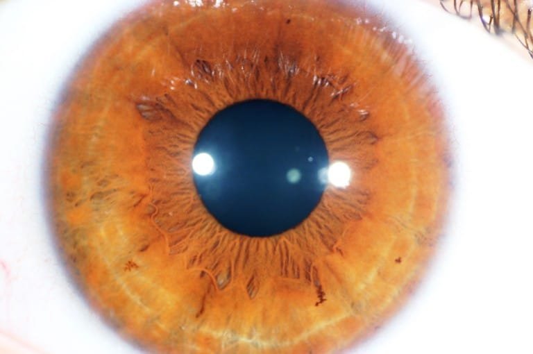
Collagen trabeculae that surround the border of the crypts can be seen in blue irises.
YELLOW RING AROUND IRIS OF EYE SERIES
The crypts of Fuchs are a series of openings located on either side of the collarette that allow the stroma and deeper iris tissues to be bathed in aqueous humor.Many fish have neither, and, as a result, their irises are unable to dilate and contract, so that the pupil always remains of a fixed size. The muscle cells of the iris are smooth muscle in mammals and amphibians, but are striated muscle in reptiles (including birds). The root of the iris is the thinnest and most peripheral. Radial ridges extend from the periphery to the pupillary zone, to supply the iris with blood vessels. It is typically defined as the region where the sphincter muscle and dilator muscle overlap. The collarette is a vestige of the coating of the embryonic pupil. The collarette is the thickest region of the iris, separating the pupillary portion from the ciliary portion. The ciliary zone is the rest of the iris that extends to its origin at the ciliary body.The pupillary zone is the inner region whose edge forms the boundary of the pupil.The iris is divided into two major regions: The iris along with the anterior ciliary body provide a secondary pathway for aqueous humour to drain from the eye. Just in front of the root of the iris is the region referred to as the trabecular meshwork, through which the aqueous humour constantly drains out of the eye, with the result that diseases of the iris often have important effects on intraocular pressure and indirectly on vision. The iris and ciliary body together are known as the anterior uvea. The outer edge of the iris, known as the root, is attached to the sclera and the anterior ciliary body. The high pigment content blocks light from passing through the iris to the retina, restricting it to the pupil. This anterior surface projects as the dilator muscles. The back surface is covered by a heavily pigmented epithelial layer that is two cells thick (the iris pigment epithelium), but the front surface has no epithelium. The constricting muscle is located on the inner border. The outer border of the iris does not change size. The pupil's diameter, and thus the inner border of the iris, changes size when constricting or dilating. The sphincter pupillae is the opposing muscle of the dilator pupillae. The iris (brown coloured portion of the eye) controls the size of the pupil by contracting the sphincter pupillae and dilator pupillae muscles The stroma is connected to a sphincter muscle ( sphincter pupillae), which contracts the pupil in a circular motion, and a set of dilator muscles ( dilator pupillae), which pull the iris radially to enlarge the pupil, pulling it in folds. The iris consists of two layers: the front pigmented fibrovascular layer known as a stroma and, beneath the stroma, pigmented epithelial cells.

YELLOW RING AROUND IRIS OF EYE PLUS
The word "iris" is derived from the Greek word for " rainbow", also its goddess plus messenger of the gods in the Iliad, because of the many colours of this eye part. In optical terms, the pupil is the eye's aperture, while the iris is the diaphragm. How can you tell if your cat is feeling feverish? Find out next.In humans and most mammals and birds, the iris ( PL: irides or irises) is a thin, annular structure in the eye, responsible for controlling the diameter and size of the pupil, and thus the amount of light reaching the retina. If you notice any type of injury or abnormality with your cat's eyes, take him or her to the vet immediately to get it checked out.Ĭats run hotter than humans, but that doesn't mean that they can't get fevers. One of the ways cats announce that they don't feel well is when their third eyelids are up - that is, they've moved partially across the eyeball. Again, it's something you rarely notice unless there's a problem. The third eyelid, or nictitating membrane, appears when your cat blinks or closes his eyes this wonderful adaptation moves from the inside corner of the eye to cover the front surface of the eyeball. You usually don't notice the conjunctiva unless it swells up, in which case it may protrude from under the eyelid, giving the eye a "meaty" appearance.

Yellow or "bloodshot" sclera, ulcers or splotches of color, and signs of damage (like scrapes or bruises) are indicators of trouble.Ĭonjunctiva is the pink, fleshy stuff under the eyelids that helps hold the eye in place. Obviously, this should be white (perhaps with some small blood vessels visible). The "white" of the eye is officially known as the sclera. Old cats' irises may degenerate and take on dark, freckle-like spots, but this isn’t always a sign of trouble. It's not unusual for the iris to change with age. If you notice changes in your cat's iris or the appearance of splotches of other colors, contact your vet. Occasionally, a cat will be "odd-eyed" each eye is a different color. Cats usually have some variety of green, yellow or blue eyes.


 0 kommentar(er)
